by
John R. Fischer, Senior Reporter | March 12, 2024
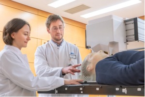
Maria Martisikova, the project leader from Heidelberg University Hospital and German Cancer Research Center (DKFZ), and DKFZ researcher Laurent Kelleter (Photo courtesy of Heidelberg University Hospital / H.Schroeder)
For the first time, German researchers are testing a new imaging solution on patients to see if they can mitigate the extent of damage that ion beam radiotherapy has on surrounding healthy tissues.
According to the German National Center for Tumor Diseases (NCT), the German Cancer Research Center (DKFZ), and the Heidelberg Ion Beam Therapy Center (HIT) at Heidelberg University Hospital, ion beams can be directed to the exact depth inside the human head necessary for most effectively irradiating tumors. The problem is that the radiation from these beam particles also affects surrounding healthy tissue and risks damaging the optic nerve or a patient’s memory when applied to the brain.
With current technologies unable to precisely target ion beams to tumors, the three organizations are hoping that a new device developed by Czech company ADVAMed will change this and are assessing its potential in head and neck tumors. The ADVACAM device tracks secondary particles created when ions pass through the head, allowing it to improve the direction of these beams. It also accounts for changes in the brain following CT "map" scans, allowing clinicians to adjust treatment.
"Our cameras can register every charged particle of secondary radiation emitted from the patient's body. It's like watching balls scattered by a billiard shot. If the balls bounce as expected according to the CT image, we can be sure we are targeting correctly. Otherwise, it's clear that the 'map' no longer applies. Then it is necessary to replan the treatment," said Lukáš Marek, of ADVACAM, in a statement.
The solution includes a small imepix3 pixel detector, a general-purpose integrated circuit developed at CERN, to closely monitor head and neck tumors to target them more easily during treatment and limit the treatment’s side effects. Its design is meant to emulate a camera, capturing and counting each individual particle, allowing for high-resolution, high-contrast, noiseless images.
The technology was originally developed for detectors used in fundamental physics research, and testing like this shows its application can be extended to other fields, including healthcare.
In the first phase, the data the ADVACAM device collects could allow clinicians to stop and replan irradiation sessions when necessary. The researchers are aiming to eventually help develop a system that can correct the path of the ion beam in real time.
“It will allow us to reduce the overall irradiated volume of tissue, saving healthy tissue and reducing the side effects of radiotherapy. We will also be able to apply higher doses of radiation to the tumor" said Mária Martišíková.
Back to HCB News