by
John R. Fischer, Senior Reporter | October 14, 2019
Ultrasound and cone beam CT imaging can
ensure greater precision of radiation delivered to
treat cervical cancer
Combining CT scans with other forms of medical imaging may reduce the amount of healthy tissue exposed to radiation in radiotherapy procedures and subsequently minimize side effects.
That’s what researchers at the Institute of Cancer Research and the Royal Marsden NHS Foundation Trust are claiming. Divided into two groups, the researchers at both tested two imaging techniques that combine CT with ultrasound and SPECT to help form more efficient treatment plans for cases of cervical and lung cancers, respectively. They expect the combinations to create greater potential for more precise delivery of radiotherapy.
CT is often employed to determine how much tissue will be exposed to radiation, with a treatment plan drawn up and depicting margins around the tumor to ensure the whole mass is treated. These margins, however, expose healthy tissue to damaging radiation, leading to patients experiencing side effects after treatment.


Ad Statistics
Times Displayed: 364973
Times Visited: 6957 Quality remanufactured Certified Centrifuges at Great prices! Fully warranted and backed by a company you can trust! Call or click for a free quote today! www.Centrifugestore.com 800-457-7576
This can especially be true when devising treatment plans for cervical cancer, which often include wide margins around the tumor to account for natural movements of the bladder and bowel that cause the tumor to shift position. In addition to side effects, women may not be able to complete treatment as a result.
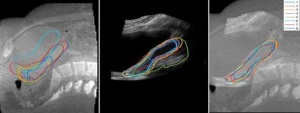
To address this, the researchers propose using ultrasound, which is quick and effective for producing high-quality images, with cone beam CT, which can produce images that suit each patient’s anatomy each day when planning radiotherapy. The authors also point out that ultrasound is relatively cheap to perform and simple to incorporate into current radiotherapy practices.
Applied to 11 patients, the two modalities together produced more detailed images than either scan alone. The scans from each complemented one another by showing regions that were not visible on a single view, enabling clinicians to better outline the positions of the uterus and improve the precision of radiotherapy planning.
"Patients with cervical cancer receive radiation to the uterus and cervix. The position and shape of the uterus can vary greatly day to day and it is important we are able to visualize them for therapy," Dr. Emma Harris, leader of the Imaging for Radiotherapy Adaptation Team at the Institute of Cancer Research, told HCB News. "The uterus and cervix are easily visualized using ultrasound and therefore, are good candidate sites on which to test this technique. Other tumour sites that will benefit from this technology are prostate and liver. We are continuing to develop these techniques for cervix, liver and prostate."