by
John R. Fischer, Senior Reporter | May 01, 2019
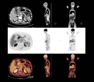
A new technique generates higher-
quality PET images more than 100
times faster than conventional methods
A team of New York-based clinicians has developed a new technique for generating PET images of higher quality more than 100 times faster than conventional methods.
Researchers at Memorial Sloan Kettering Cancer Center have harnessed the power of artificial intelligence to train a convolutional neural network to convert raw PET data into images. They call their method DeepPET.
“No one has done PET imaging in this way before,” said medical physics researcher Dr. Ida Häggström in a statement, adding that “the gain we’ve seen in reconstruction speed and image quality should lead to more efficient image evaluation and more reliable diagnoses and treatment decisions, ultimately leading to improved care for our patients.”


Ad Statistics
Times Displayed: 365483
Times Visited: 7057 Quality remanufactured Certified Centrifuges at Great prices! Fully warranted and backed by a company you can trust! Call or click for a free quote today! www.Centrifugestore.com 800-457-7576
The technique involves teaching the computer system to identify features in the training data and apply the knowledge to data never before seen, enabling it to complete tasks such as classifying malignant lesions, predicting outcomes of treatment, or interpreting medical charts.
While converting data into PET images traditionally takes a long time and may generate pictures that are not always clear, the researchers trained the computer network using large amounts of PET data, along with the associated images. They also taught it about the scanner’s physical and statistical characteristics, and how typical PET images looked, so as to eliminate the conventional repeating process associated with PET generation and replace it with production of an image by a single, fast computation.
The system is currently under preparation for clinical testing. “By combining that expertise with the state-of-the-art computational resources that are available here, we have a great opportunity to have a direct clinical impact,” said Häggström.
Research was funded in part through a National Institutes of Health/National Cancer Institute grant.
The findings were published in the May 2019 issue of
Medical Image Analysis.