Researchers have found that immune cells play a role in the repair of the meninges following the occurrence of mild traumatic brain injury
Different immune cells play key roles in the recovery process of the brain following mild traumatic brain injury (mTBI), according to a new study.
Researchers at the National Institutes of Health, drawing from observations of human brain MR scans observed, in real time, different immune cells in the brains of mice carrying out carefully timed tasks to fix damage incurred by the meninges, concluding that such findings may enable a better understanding of the repair process in humans and help identify potential targets for therapy to improve patient outcomes.
“Our studies both in mice and humans show that blood vessels in brain lining have the ability to repair, following mild traumatic brain injuries,” Dr. Dorian McGavern, a scientist at the NIH’s National Institute of Neurological Disorders and Stroke, told HCB News. “We have also shown that peripheral immune cells recruited from the blood play an important role in the reparative process.”
MIT labs, experts in Multi-Vendor component level repair of: MRI Coils, RF amplifiers, Gradient Amplifiers Contrast Media Injectors. System repairs, sub-assembly repairs, component level repairs, refurbish/calibrate. info@mitlabsusa.com/+1 (305) 470-8013
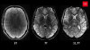
The study was inspired by MR scans taken of adult human patients who experienced a concussion or mTBI. Damaged blood vessels in the meninges were found severed in half with mTBI, appearing in the form of a vascular dye leaking out of the damaged vessels on the scans.
Though leaks in most patients were repaired within 20 days, leakage still showed up in scans for 17 percent three months following their injuries, indicating ongoing meningeal damage.
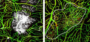
Researchers then employed intravital two-photon laser scanning microscopy to film in real time the injury and repair process, assessing the pathology and immune cell dynamics in the brain lining of the mice for up to one week after an injury.
Their findings showed that blood-based immune cells known as inflammatory monocytes center the core of the damaged tissue within the first day of the injury to clear out dead cells, and are assisted a few days later by a different type of blood monocyte which works around the lesion edge to help rebuild damaged blood vessels over the course of one week.
Further observations showed that the actions of neither overlapped the other, or one took over when the activities of another were blocked.
The scientists also recorded the impact that the timing of a second head injury has on the repair process, finding in mice that the occurrence of a second injury within one day of the first TBI caused additional inflammation, with the blood vessels remaining unfixed during the repair.