by
Lauren Dubinsky, Senior Reporter | April 12, 2018
Cardiac, breast, chest, MSK
imaging tools to come
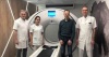
A team of researchers at the Ohio State University Wexner Medical Center (OSUMC) have developed an AI tool that can detect critical findings on head CT, such as hemorrhage and stroke.
“Currently, we are planning to release the ‘AI for Head CT algorithms’ for beta testing here at OSUMC,” Barbaros Selnur Erdal, assistant chief of the division of medical imaging informatics at the medical center, told HCB News. “AI for cardiac imaging for evaluation of coronary artery disease is next in line.”
The team used over 2,500 representative images to train the tool. It was designed to analyze the imaging dataset, detect images with critical findings and send them to the top of the radiologist’s worklist.


Ad Statistics
Times Displayed: 173767
Times Visited: 3176 For those who need to move fast and expand clinical capabilities -- and would love new equipment -- the uCT 550 Advance offers a new fully configured 80-slice CT in up to 2 weeks with routine maintenance and parts and Software Upgrades for Life™ included.
To date, the team has evaluated the performance of the tool for detecting hemorrhage, mass effect, or hydrocephalus (HMH), as well as suspect acute infarct (SAI) in non-contrast head CT exams. The results were published in
Radiology in December.
They found that when detecting HMH, the tool showed 90 percent sensitivity and 85 percent specificity, and when detecting SAI, achieved 62 percent sensitivity and 96 percent specificity. More research is needed to determine if it can independently screen non-contrast head CT exams and notify the interpreting radiologist of critical findings.
“If AI is used appropriately, it has the potential to enable the radiologist to be more efficient,” said Erdal. “Depending on the use case, AI has the potential to enable the radiologist to be more quantitative and objective on disease-specific measurements.”
He added that in addition to the cardiac AI tool, his team is also currently developing various breast imaging, chest imaging and musculoskeletal imaging applications.