by
Gus Iversen, Editor in Chief | April 23, 2018
At Brigham and Women's, surgeons
bring the iMRI to the patient rather
than the other way around
From the April 2018 issue of HealthCare Business News magazine
For decades, Brigham and Women’s Hospital in Boston has been making innovative strides across a variety of specialties to constantly improve patient outcomes and levels of care for the industry. Much of its surgical innovation is represented in a particular area of the hospital – the Advanced Multimodality Image Guided Operating (AMIGO) Suite.
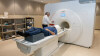
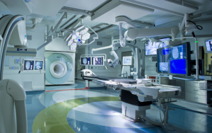
The AMIGO Suite is a state-of-the-art, custom-designed surgical research environment that houses a complete array of advanced imaging equipment and interventional surgical systems including intraoperative magnetic resonance imaging (iMRI) and intraoperative computed tomography (iCT), positron emission tomography (PET), surgical navigation systems, C-Arm X-Ray, 3-D ultrasound and a near-infrared imaging system. This collection of intraoperative and hybrid OR solutions is a one-of-a-kind suite that was developed in 2011 and in it they have treated nearly 2,000 patients.
Their innovation goes back decades earlier, however, as one aspect of the suite has held its legacy at Brigham and Women’s – intraoperative magnetic resonance imaging (iMRI).


Ad Statistics
Times Displayed: 46091
Times Visited: 1397 MIT labs, experts in Multi-Vendor component level repair of: MRI Coils, RF amplifiers, Gradient Amplifiers Contrast Media Injectors. System repairs, sub-assembly repairs, component level repairs, refurbish/calibrate. info@mitlabsusa.com/+1 (305) 470-8013
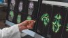
Brigham and Women’s was one of the first hospitals in the world to introduce iMRI, allowing their surgeons to access advanced anatomical imaging detail while the patient is on the operating table. Alexandra Golby, M.D., the director of image-guided neurosurgery at Brigham and Women’s, has been with the hospital for more than 15 years and has had a front row seat and contributive role in their early-adopting and forward-thinking approaches.
According to Dr. Golby, “intraoperative MRI is about being equipped with the best possible information with which to perform, optimize and personalize surgery. It’s about information, the right information, at the right time, in the right place.”
Intraoperative MRI: Lowering risks and enhancing precision
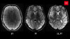
Intraoperative MRI was first introduced at Brigham and Women’s in the early 1990s to achieve better resection rates in low grade gliomas – brain tumors that are difficult to distinguish from surrounding tissue with the naked eye. Without intraoperative imaging to guide surgeons throughout procedures, the risk of a subtotal resection, or even damage to healthy tissue, runs much higher. Intraoperative MRI offers greater precision during surgery.
When the AMIGO Suite was developed in 2011, Brigham and Women’s worked with IMRIS — who offers the world's only
moving intraoperative MR solution — to raise the level of care even higher. The system allows surgeons to bring the iMRI to the patient, rather than the other way around to avoid the inherent risks of moving a patient during surgery. Lowering risks and improving surgical precision has led to increasingly positive patient outcomes, with less need for follow-up surgeries.