by
Lauren Dubinsky, Senior Reporter | November 20, 2017
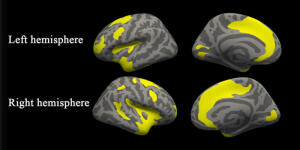
Significant cortical thickness differences
among the three groups
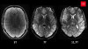
A new study published in Radiology used MR imaging to break new ground in depression and anxiety research.
“The consistent structural differences in the two patient groups may contribute to the broad spectrum of emotional, cognitive and behavioral disturbances observed in major depressive disorder patients and social anxiety disorder patients,” Dr. Youjin Zhao of Sichuan University in China, told HCB News.
Zhao and her team had 37 MDD patients, 24 SAD patients and 41 healthy controls undergo MR exams in order to spot any alterations in the brain’s gray matter. They focused the research on the thickness of the cortex, which is the principle part of the brain.


Ad Statistics
Times Displayed: 172926
Times Visited: 3140 For those who need to move fast and expand clinical capabilities -- and would love new equipment -- the uCT 550 Advance offers a new fully configured 80-slice CT in up to 2 weeks with routine maintenance and parts and Software Upgrades for Life™ included.
The imaging revealed that both the MDD and SAD patients had gray matter abnormalities in the salience and dorsal attention networks, compared to the healthy controls. The salience network determines the stimuli that deserve our attention and the dorsal attention network is associated with focus and attentiveness.
“Depression and anxiety respond to the same treatment strategies; thus, it has been suggested that they share a similar etiology, said Zhao. “There have also been claims that a similar neural, presumably computational, architecture mediates mood and anxiety symptoms.”
Both MDD and SAD patients, relative to health controls, showed cortical thickening in the brain region that’s vital to perception and self-awareness. The team thinks that could reflect a compensatory mechanism related to inflammation or be the result of continuous coping efforts and emotion regulation attempts by the patients.
They also found disorder-specific involvement of the brain’s fear circuitry in the SAD patients and involvement of the visual recognition network in patients with MDD. They hypothesized that alterations within the visual recognition network could be associated with impaired selective attention and working memory in MDD.
Zhao concluded that this study provides preliminary evidence of common and specific gray matter changes in MDD and SAD patients. She added that future studies that involve larger sample sizes and leverage machine learning technology may further aid the diagnostic and prognostic value of structural MR.