New fMRI research may help reveal susceptibility to depression in children
January 25, 2016
Alzheimers/Neurology
MRI
Pediatrics
Risk Management

By Gail Kalinoski, Contributing Reporter
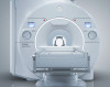
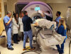
Using functional magnetic resonance imaging (fMRI) on two groups of children ages 8 to 14, researchers at the Massachusetts Institute of Technology (MIT) and Harvard Medical School have found such brain scans may be able to determine whether young people are at risk of developing depression when they are older. The researchers hope the information in their study could lead to tools that could be used for early detection, intervention and treatment.
The study, published in the journal Biological Psychiatry, is important because an estimated 2.8 million adolescents aged 12 to 17 in the United States had at least one major depressive episode in 2014, roughly 11.4 percent of the U.S. population, according to statistics from the National Institute of Mental Health. That same year, about 15.7 million American adults also had at least one major episode.


 Experts say early intervention is important because one depressive episode is likely to lead to more.
“If you can avoid that first bout, maybe it would put the person on a different trajectory,” John Gabrieli, an author of the study and professor of brain and cognitive sciences at MIT, told MIT News.
“We’d like to develop the tools to be able to identify people at true risk, independent of why they got there, with the ultimate goal of maybe intervening early and not waiting for depression to strike the person,” Gabrieli added.
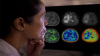
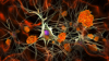
The study found distinctive brain differences in children considered high risk because of depression in their families. Previous research using imaging scans had found two brain regions – the subgenual anterior cingulate cortex and the amygdala – often showed abnormal activity in depressed patients. But it wasn’t clear if the differences were caused by the depression or if the depressive episode changed the brain.
Gabrieli and his team used brain scans of children who were not diagnosed as depressed but had a parent with depression because statistics show those children are three times more likely to eventually become depressed, often by age 15. The researchers studied 27 high-risk children ages 8 to 14 and compared them to 16 children with no family history of depression.
In the at-risk children, they found similar patterns to those in adults with depression leading them to believe the differences are there before depression occurs and may contribute to it.
More research will be done to track those at-risk children in the study to see whether early treatment might prevent later depression, or how some could avoid depression without treatment.
Other studies using fMRI have been done involving children and depression. A 2013 study by researchers at Washington University School of Medicine in St. Louis found differences in the amygdala of young children with depression. In the study, 54 children between the ages of 4 and 6 were tracked. Of those, 23 had been diagnosed with depression.
The fMRI data showed that children with depression had elevated activity in the amygdala, according to the study published in the July 2013 issue of the Journal of the American Academy of Child & Adolescent Psychiatry. This group of researchers also said they hoped the results of the fMRIs would lead to new ways to identify and treat young children suffering from the condition.
Using functional magnetic resonance imaging (fMRI) on two groups of children ages 8 to 14, researchers at the Massachusetts Institute of Technology (MIT) and Harvard Medical School have found such brain scans may be able to determine whether young people are at risk of developing depression when they are older. The researchers hope the information in their study could lead to tools that could be used for early detection, intervention and treatment.
The study, published in the journal Biological Psychiatry, is important because an estimated 2.8 million adolescents aged 12 to 17 in the United States had at least one major depressive episode in 2014, roughly 11.4 percent of the U.S. population, according to statistics from the National Institute of Mental Health. That same year, about 15.7 million American adults also had at least one major episode.

We repair MRI Coils, RF amplifiers, Gradient Amplifiers and Injectors.
MIT labs, experts in Multi-Vendor component level repair of: MRI Coils, RF amplifiers, Gradient Amplifiers Contrast Media Injectors. System repairs, sub-assembly repairs, component level repairs, refurbish/calibrate. info@mitlabsusa.com/+1 (305) 470-8013
“If you can avoid that first bout, maybe it would put the person on a different trajectory,” John Gabrieli, an author of the study and professor of brain and cognitive sciences at MIT, told MIT News.
“We’d like to develop the tools to be able to identify people at true risk, independent of why they got there, with the ultimate goal of maybe intervening early and not waiting for depression to strike the person,” Gabrieli added.
The study found distinctive brain differences in children considered high risk because of depression in their families. Previous research using imaging scans had found two brain regions – the subgenual anterior cingulate cortex and the amygdala – often showed abnormal activity in depressed patients. But it wasn’t clear if the differences were caused by the depression or if the depressive episode changed the brain.
Gabrieli and his team used brain scans of children who were not diagnosed as depressed but had a parent with depression because statistics show those children are three times more likely to eventually become depressed, often by age 15. The researchers studied 27 high-risk children ages 8 to 14 and compared them to 16 children with no family history of depression.
In the at-risk children, they found similar patterns to those in adults with depression leading them to believe the differences are there before depression occurs and may contribute to it.
More research will be done to track those at-risk children in the study to see whether early treatment might prevent later depression, or how some could avoid depression without treatment.
Other studies using fMRI have been done involving children and depression. A 2013 study by researchers at Washington University School of Medicine in St. Louis found differences in the amygdala of young children with depression. In the study, 54 children between the ages of 4 and 6 were tracked. Of those, 23 had been diagnosed with depression.
The fMRI data showed that children with depression had elevated activity in the amygdala, according to the study published in the July 2013 issue of the Journal of the American Academy of Child & Adolescent Psychiatry. This group of researchers also said they hoped the results of the fMRIs would lead to new ways to identify and treat young children suffering from the condition.
You Must Be Logged In To Post A CommentRegisterRegistration is Free and Easy. Enjoy the benefits of The World's Leading New & Used Medical Equipment Marketplace. Register Now! |
|